Pet owners often wish they could stay with their dog or cat for dental procedures. They wonder about the processes and what they entail. This is understandable. Knowing what therapy or treatment your pet is receiving allows you to be confident and comfortable—and at Wales Animal Clinic, not only do we want to ensure that your pet’s comfort is a priority, but we also want to ensure your comfort as well. So let’s take an inside look at one critical component of a comprehensive pet dental examination—one that many pet owners might not know about—dental X-rays.
What are dental X-rays for pets?
Dental X-rays are images of your pet’s teeth and oral cavity, taken under anesthesia with a small X-ray machine and a piece of film or a small digital sensor placed inside your pet’s mouth. Dental X-rays are often digital, which allows the veterinarian to read the image on a computer. Digital X-rays provide a high resolution image and capture more detail than traditional film, and they take less time because they do not require development.
A precise image is necessary for diagnosis, and this requires your pet to be still, as well as carefully positioned. To obtain good results, pets must undergo general anesthesia for dental X-rays and cleaning. After your pet’s entire mouth is imaged—that’s X-rays of up to 42 teeth for dogs and 30 for cats—our veterinarians can review the X-rays, perform an oral exam, and form a treatment plan.
Dental X-rays—telling the whole story of pet oral health
In veterinary dentistry, the whole tooth—as well as the surrounding gum tissue and bone—must be considered when evaluating your pet’s oral health and viability. The crown of your pet’s tooth may appear white and flawless, while under the gumline the true story is revealed—courtesy of the dental X-ray.
What pet dental X-rays reveal
- Periodontal disease — This is an infection and inflammation around the tooth, caused by plaque bacteria. Periodontal disease will appear on an X-ray as deterioration and bone loss around a tooth and or as a damaged or nonexistent periodontal ligament, which holds the tooth in place. Gingivitis—inflammation of the gums—may be visible at the tooth surface.
- Unerupted teeth — Often found by accident, hidden adult teeth that never broke through the gum line may show up on X-rays.
- Resorptive lesions — Most often seen in cats, resorptive lesions are a painful condition that occurs when a tooth breaks down from the outside, beginning at the surface of the root and then upward toward the crown. On an X-ray, the roots of a tooth severely affected by resorptive lesions may be partially or completely absent, with only the crown remaining. Redness may be present along the gumline, as the inflamed tissue tries to fill the space left behind by the eroding tooth.
- Fractured teeth — Some fractures are obvious above the gumline, but X-rays can show fractured roots, which require the tooth to be removed.
- Jaw fractures — Old or fresh jaw fractures—often seen in smaller dogs and cats—can be found on a dental X-ray. Our veterinarians can also examine the X-ray for signs of cancer, which, along with trauma, is a common reason for jaw fracture.
- Cancer — Dental X-rays can be used to determine if an oral tumor has eroded or invaded bone, affecting the prognosis and treatment.
- Root fragments — The remnants of past dental work can be another finding on dental X-rays. Most often you’ll see roots left behind from previous extractions.
- Post-op X-rays — Before closing the gum tissue over extractions, the veterinarian will take a final X-ray to confirm that all pieces of the root have been removed from the tooth socket. Pieces of root left behind can create a prime location for painful infection. After the X-ray, the site is sutured closed to promote gum healing and protect the empty socket.
Safety of pet dental X-rays
Many pet owners wonder if taking dental X-rays is safe for pets. The answer is yes. Dental X-ray beams are highly focused, or collimated, which prevents dangerous “scatter” radiation. The X-rays, themselves, are one-third the amount of radiation a pet receives from a standard X-ray, making it safe to take images from multiple angles and directions.
How pet dental X-rays save you money
On a dental examination estimate, X-rays look like another line item in a long list. Many times, pet owners will decide to skip the X-rays to save money. However, over the course of your pet’s lifetime, dental X-rays will actually reduce veterinary costs. Here are two examples:
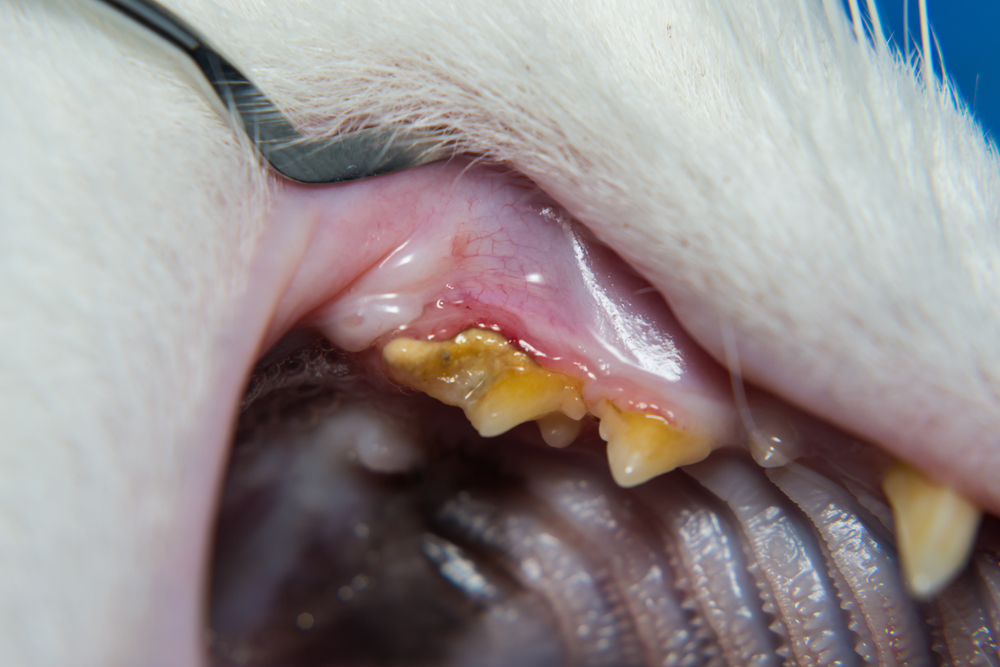
- Early intervention — Early periodontal disease is visible along a dog’s lower premolars. The veterinarian reviews the X-ray and concludes the nearby teeth do not show signs of damage. She applies an oral antibiotic in the pocket of the gumline to clear the bacteria and save the teeth, instead of extracting them. At the dog’s next dental cleaning, no signs of periodontal disease appear.
- Preventive care — A senior dog has multiple teeth extracted after dental X-rays reveal root erosion. His oral health significantly improves, and he does not develop heart, liver, or kidney failure from dental disease, saving his owner thousands of dollars and heartache.
The importance of pet oral health
Maintaining oral health means a long, healthy life for your pet and requires commitment and dedication. Many great home care options are available for preventing plaque and tartar, but nothing compares to a comprehensive oral exam and dental cleaning at Wales Animal Clinic. Call us to schedule a dental consultation for your pet.



Leave A Comment